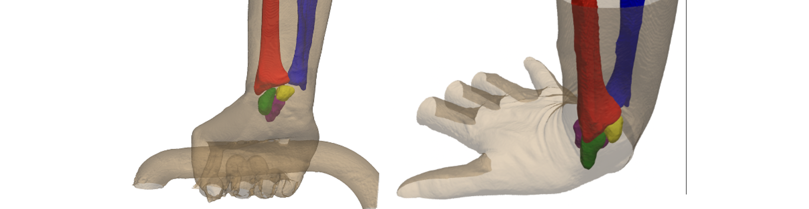
Assessing Carpal Alignment and Kinematics Using WBCT
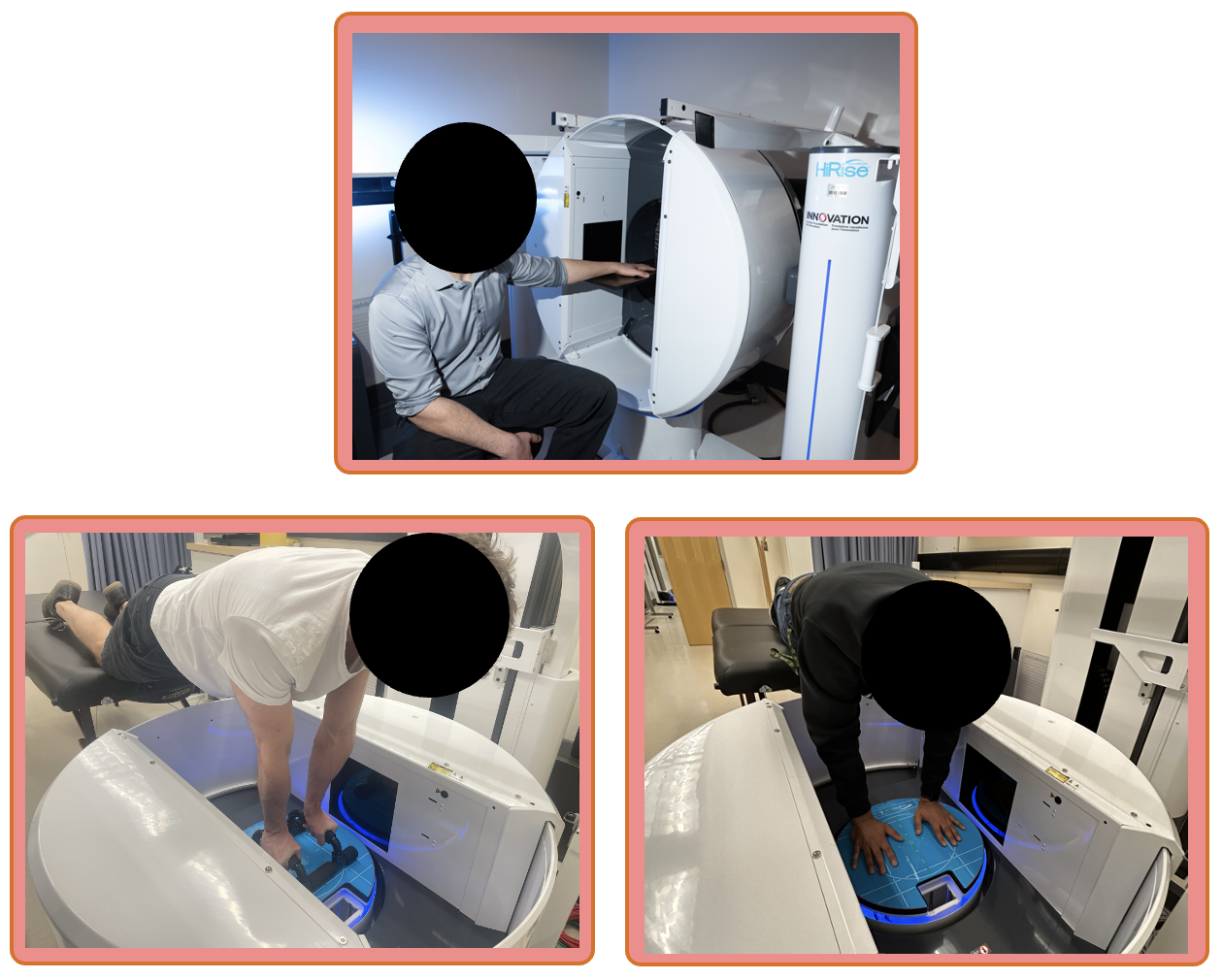
Research Images
Methodology and Goals
When patients are in the intensive care unit (ICU), their muscles can weaken, causing long-lasting problems with muscle strength and daily activities. We do not fully understand why this happens, and measuring muscle strength in the ICU is challenging. The goal of our CIHR-funded project is to figure out how, when and why this muscle weakness occurs and how it affects people after leaving the ICU. We will use computed tomography (CT) scans that were already acquired while patients were in the ICU to:
1) Establish the risk factors and time course of muscle atrophy in the ICU. Using the multiple clinically acquired CT scans (often > 2 per patient), we have a novel opportunity to characterize the rate of abdominal muscle atrophy and factors associated with muscle atrophy during the ICU stay.
2) Determine how ICU-induced muscle loss is related to long-term recovery and physical function. Our novel data measuring muscle cross-sectional area indicates that ICU-induced muscle loss is associated with ICU mortality. However, whether CT assessment of ICU-associated muscle atrophy is related to long-term outcomes is unknown.
3) Advance CT imaging measures of muscle fat infiltration and fibrosis using internal calibration to measure muscle density.
In addition to atrophy, muscle infiltration with adipose and fibrosis is frequently implicated in loss of muscle function. Whether clinical-quality CT scans can accurately capture muscle density changes is unknown.
This research will help us understand why muscles weaken without needing an invasive biopsy. Using CT scans, we can understand when, how and in whom muscles weaken in the ICU to better target rehabilitation strategies.